Nuclear Scintigraphy
Nuclear scintigraphy uses very small, tracer amounts of radioactive molecules to diagnose diseases involving bone, soft tissues and vessels. We can attach these molecules to agents that bind to bone lesions, soft tissue tumors and sites of infection. This very sensitive technique can often diagnose diseases not visible with other imaging methods.
The radiotracer eventually collects in the area of the body being examined, where it gives off energy in the form of gamma rays. This energy is detected by a device called a gamma camera. These devices work together with a computer to measure the amount of radiotracer absorbed by your body and to produce special pictures offering details on both the structure and function of organs and other internal body parts.
Image One

Nuclear scintigraphy is most commonly used for bone scans in horses. A radiopharmaceutical is given IV that binds to areas of exposed hydroxyappetite in the bone. This radiopharmaceutical gives off gamma rays that are detected by a gamma camera.
Bone scans are useful for horses with multiple limb lameness, subtle lameness or lameness of the proximal limb, back or pelvis.
The spatial resolution is poor but bone scans are used to locate areas of disease and direct further imaging.
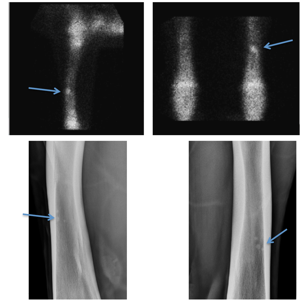
Joints and physes normally have more uptake than the rest of the bone. In this example there is focal radiopharmaceutical uptake in the mid-diaphysis of the left radius (blue arrow). This area of uptake corresponds to three small focal enostoses or areas of new bone within the medullary cavity.
Image Two

The horse is giving off radiation and the gamma camera is moved around the horse to obtain different views.
These are images of the pelvis with the camera above the horse so that the lumbar spine is at the top of each image and the wings of the ilium extend to either side.
There is asymmetry in the pelvis of this horse with severe focal radiopharmaceutical uptake of the right tuber sacrale or point of the croup. This represents a fracture in this patient.
