Improving Animal Vision
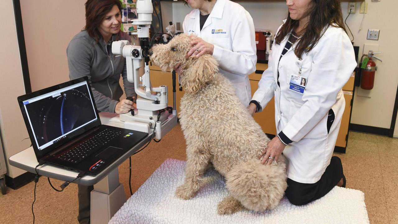
It’s fitting that Dr. Sara Thomasy is an ophthalmologist. Her eyes light up when she talks about the breakthroughs being made at UC Davis thanks to recent acquisitions of state-of-the-art imaging equipment. Eight new pieces of imaging equipment and one new piece of laboratory technology, made possible by grants from the Center for Companion Animal Health (CCAH), now allow the Ophthalmology Service to provide new levels of care.
“All of these have advanced the way we practice,” said Thomasy. “As an example, we just examined a dog with an injury that previously would’ve never been discovered. We struggled for weeks to determine why this patient had so much corneal swelling. When we did imaging with the spectralis, we saw that a layer of the cornea was detached.”
The spectralis unit provides detailed cross-sectional imaging of the cornea, anterior chamber, iris, retina and optic nerve and is commonly utilized in physician-based ophthalmology for clinical research and patient care.
Human and animal eye similarities
With striking similarities between human and animal eyes, veterinary ophthalmologists at the school are able to collaborate with their human medicine counterparts at UC Davis Health. Ophthalmology in veterinary medicine is arguably the closest resembling specialty service to human medicine. Understanding there is much to be learned from both sides, researchers are utilizing new technologies that could lead to shared breakthroughs.
CCAH provided equipment funds to acquire an electroporator, a piece of laboratory equipment that allows genetic modification of cells in culture dishes.
“Through the use of this technology, we can better understand the cell signaling interactions that lead to scarring of the eye,” said Dr. Brian Leonard, an assistant professional researcher in comparative ophthalmology. “With this knowledge, we are able to develop novel therapeutics to improve the surgical outcome in veterinary and human patients.”
“We strive to operate our Ophthalmology Service at a similar level of practice as human medicine,” Thomasy said. “Sometimes the goals are a bit different because our patients don’t have to read or drive, but that doesn’t diminish the levels we are reaching in veterinary medicine.”
Thomasy recently collaborated with an ophthalmologist from UC Davis Health to diagnose epithelial downgrowth in a dog, a rare, aggressive ocular complication that can cause blindness. Because of the new imaging modalities utilized, they were able to find an open wound in the cornea and correct it with surgery. This dog had already lost one eye prior so saving the remaining eye was crucial.
“Our chances of success with this surgery were definitely higher because of the imaging capabilities of our new equipment,” beamed Thomasy.
New discoveries through clinical trials
The Ophthalmology Service has numerous clinical trials for which the equipment is a vital component of the study. The instrumentation allows the team to critically evaluate structures within the eye in a greatly magnified manner, photographically document the process, and monitor intricate changes occurring throughout each study. This ultimately helps guide their ability to provide the most optimal therapy for those specific diseases.
“This new instrumentation has been absolutely imperative for our researchers to run clinical trials and immediately transferring that to the clinic when new discoveries have proven successful,” said Dr. Kathryn Good, chief of the service. “The information generated by this equipment has directly benefitted our patients.”
Researchers are utilizing the imaging equipment to study three of the leading causes of blindness in dogs: sudden acquired retinal degeneration syndrome, corneal endothelial dystrophy, and glaucoma. It’s also used to advance corneal transplants. Thomasy credits a new ultrasound biomicroscope—which obtains high resolution, extremely magnified ultrasound views of structures inside of the eye for which there is no other non-invasive means to do so—with saving the sight of a golden retriever on which she performed a full thickness corneal scleral transplant, one of the rarest and most advanced procedures in veterinary ophthalmology.
“Without that ultrasound equipment, I would’ve had to remove the eye,” Thomasy states. “This technology is adding rigor to our clinical trials, and it’s helping our clients better understand their pets’ diseases.”
“We could not provide this level of care and research without the support of CCAH,” added Thomasy. “We’re now able to do imaging and patient care that is not rivaled at any other veterinary hospital. (CCAH Director) Dr. Kent’s championing of this grant program has changed everything for us in the Ophthalmology Service.”
“We’ve been fortunate to be the recipient of these grants,” Good continued. “They’re helping to fulfill our goals of preserving vision and comfort in companion animals.”
# # #
