Neurosurgery Discovers Cause of Cat’s Seizures
How Did a Tapeworm Generally Contracted Outdoors Infect an Indoor Cat?
“Case of the Month” – February 2025
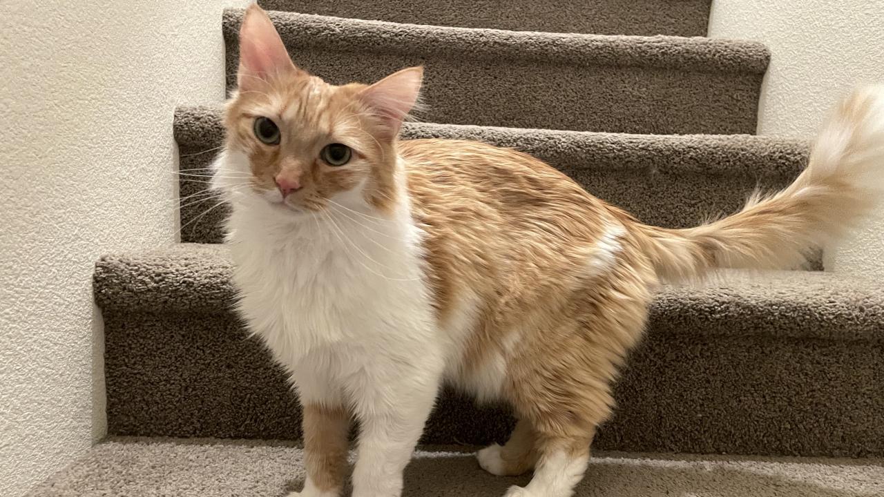
Cub, a 2-year-old orange domestic longhair cat, was far too young to be acting so lethargic last May. He was sleeping more and was less active than the other cats in the household. His owner, Grace Jesensky, also noticed his breathing was abnormal, and his primary veterinarian suspected he had asthma.
In August, Cub had a seizure, and Jesensky took him to the emergency room. A neurologic examination was unremarkable, but two more seizures followed just a few weeks later. Jesensky insisted on further testing to discover what was causing his problems.
Cub had an MRI on October 1 with Dr. Kelley Vurik, a referring neurologist at BluePearl Pet Hospital in Reno, Nevada, who found a lesion on the front temporal lobe of his brain. After performing cerebrospinal fluid analysis and common infectious disease tests, it was still uncertain what the mass in Cub’s brain truly was. Dr. Vurik then contacted the Neurology/Neurosurgery Service at the UC Davis veterinary hospital and scheduled a referral appointment for October 23. A week before that appointment, Cub had two more seizures.
“The mass on Cub’s brain was atypical for such a young cat,” said Assistant Professor Adrien Dupanloup. “Based on his MRI, we were not able to exactly identify it. Possibilities included an infectious abscess or granuloma, a tumor, or a cystic structure among other possibilities.”
Dr. Dupanloup and neurology/neurosurgery resident Dr. Kimberly Worland discussed several options with Jesensky, including surgery to obtain a biopsy with potential to remove the mass, or continuing to monitor the situation and repeat an MRI in two weeks to evaluate any change.
Jesensky chose to monitor the situation and scheduled a follow-up in November. But just the next day, Cub had a focal facial seizure, so Jesensky elected to return to UC Davis on October 29 and have Cub undergo brain surgery on October 30.
The service recently launched a neurosurgery specialty sub-service to offer a wide range of specialized neurosurgical services for complex, non-routine neurosurgical conditions like what Cub was presenting. Neurosurgeons Dr. Dupanloup and Dr. Ji-Hey Lim reviewed Cub’s MRI and determined that a transorbital approach would be the best way to remove the mass.
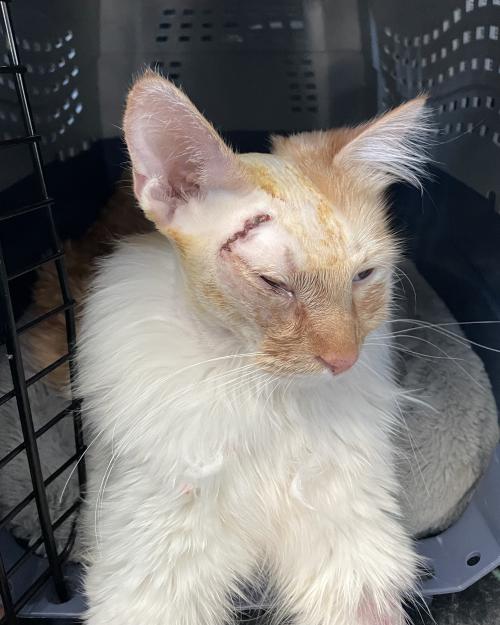
The Anesthesia Service prepared Cub for a craniotomy and continually monitored him throughout the procedure. In this operation, a small bone window was removed from Cub’s skull from behind his eye to allow the neurosurgical team of Drs. Dupanloup, Lim, Worland and fourth-year DVM student Samatha Watts to successfully remove the mass on his brain. The neurosurgeons then positioned replacement meninges (membrane layers that protect the brain) and covered the area with muscle and soft tissue to close the surgical site.
“This area is behind Cub’s eye socket that is not visible to anyone looking at him,” said Dr. Worland. “It is a rare surgical approach but was the best way to access the mass.”
“We’re really happy that we decided to go back and do the surgery immediately because when we heard the results, we knew it was only destined to get worse and grow larger,” said Jesensky.
Following surgery, Cub woke up in the Intensive Care Unit, where neurosurgeon Dr. Vishal Murthy and critical care specialist Dr. Kate Hopper monitored his neurological status and electrolytes to ensure smooth recovery from the brain procedure.
The biopsy results showed that Cub had a cerebral coenurosis – a parasitic infection of a tapeworm larvae that formed a cyst in his brain.
“Parasites in the brain are quite rare,” said Dr. Dupanloup. “Based on the morphological evaluation performed by the Clinical Pathology Service, Taenia and Echinococcus are the two potential types of tapeworms it could be.”
The definitive hosts of Taenia are wild dogs, foxes, and coyotes which usually become infected by eating raw, cyst-infected intermediate hosts like rabbits or rodents. For cats to be infected with these species, it is thought they need to ingest the tapeworm eggs in infected animal stool.
Cats are considered aberrant hosts in which the tapeworm larvae do not behave normally and migrate into abnormal tissue outside the gastrointestinal tract and form larvae cysts, like what was found in Cub’s brain. Seizures are the most common symptom of Taenia infections in the brain. This is thought to be due to inflammation associated with dying larvae in the cyst, of which Cub’s biopsies showed evidence.
However, Cub is a 100% indoor cat, so where would he have ingested the tapeworm eggs?
“At first, we thought maybe he got it from contaminated food,” Jesensky said. “But all the cats in our house share everything, and we don’t believe the others to be infected. So, now the theory is that Cub contracted the parasite in utero, and it’s lived in his system ever since.”
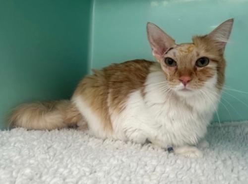
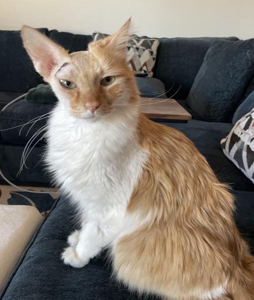
Following surgery, Cub was treated for a week with a combination of medications to combat worm and parasite infections. He is also taking anti-seizure medication. Jesensky reports that his seizures have stopped, and his energy is back to normal levels for a young cat.
“It is difficult to predict the long-term prognosis of Cub because the literature is so scarce in cats with this disease,” said Dr. Dupanloup. “However, we are pleased with his progress since the surgery and grateful for the entire team's dedication to providing him with the best possible quality of life.”
# # #