Emergency Exploratory Surgery Helps Determine Cause of Disease and Saves Kitten
“Case of the Month” – January 2022
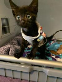
Miso, a 1-month-old kitten, and his two siblings were discovered by a Good Samaritan and brought to the UC Davis veterinary hospital’s Emergency Room on a Friday night after the local shelter was closed. They all appeared to be having trouble breathing, and the ER’s critical care specialists diagnosed Miso and his siblings with an upper respiratory tract infection, which is common in young kittens.
The following day, Miso and his siblings were officially transferred into the custody of the Yolo County SPCA, which has worked with the school to treat hundreds of kittens over the years. Dr. Karen Vernau, faculty advisor for the Orphan Kitten Project, maintained supportive care of Miso and his siblings following release from the ER until Dr. Megan Mangini, a resident in the Anesthesia Service, took over their care and fostered them a few days later.

After a few weeks, Miso’s breathing troubles suddenly increased, and he was brought back to the ER where he was cared for by Dr. Jamie Burkitt. X-rays showed a potential hernia of his diaphragm thought to be allowing his abdominal organs to fill up the chest and make breathing and eating difficult. Miso’s care was transferred to the Soft Tissue Surgery Service for exploratory surgery into his abdomen to investigate his diaphragm and repair it if necessary. Surgical resident Dr. Amy Downey and faculty surgeon Dr. Michelle Giuffrida found no hernia in the diaphragm, although his spleen was swollen with abnormal growths and had to be removed. Tissue samples of the spleen were submitted to the Anatomic Pathology Service to help understand his disease condition.
The surgeons extended Miso’s surgery into his chest cavity and discovered a build-up of very thick fluid around the left lung (pyothorax) along with severe build-up of a protein formation (fibrin) overlying his lung lobes. Fibrin build-up causes the blood to clot and impedes its flow. The lungs were not inflating properly due to the abnormal material in the chest. Samples from this area of Miso’s thoracic cavity were also collected and sent to pathology to help guide the veterinarians in their antimicrobial therapy. Finally, surgeons placed a chest tube and a gastric feeding tube in Miso for post operative care.
Dr. Mangini handled the anesthesia during Miso’s surgery and maintained him in a stable condition throughout the procedure. He recovered without complication and was maintained on IV pain medications, supportive care, and was closely monitored for any signs of post-operative or post-anesthetic complications. He started eating on his own the night after his surgery.

Pathology results showed Miso’s infection to be caused by the Streptococcus canis bacteria, which can affect very young and very old animals, as well as spread rapidly through densely populated areas, like shelters. The development of disease can occur rapidly, and respiratory infections are one of the most common symptoms. Untreated, the spread of this bacteria can quickly become fatal.
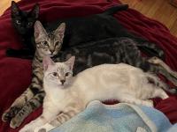
Following four days of hospitalization and a more targeted approach to treat the bacterial infection, Miso was breathing on his own and was well enough for discharge. Six weeks after his surgery, Miso was examined and was assessed to be healthy, and ready to be neutered. Miso and his brother Lucas and sister Stevie were all spayed and neutered by veterinary students and veterinarians with the hospital’s Community Surgery Service. Two months after the surgery, Miso and his two siblings were adopted by Dr. Mangini.
# # #
